01/2022, Review , 009-020


Changes in serum testosterone during the menstrual cycle – an integrative systematic review of published literature

Abstract

The effect of sex hormones in the pathogenesis of many diseases such as rheumatoid arthritis, systemic lupus erythematosus and asthma has been highlighted in addition to their role in reproductive physiology. Among the sex hormones, the effects of testosterone (T) in females constitute an important area of research. The present review critically evaluates the published literature on serial T measurements during the menstrual cycle (MC) in healthy females. Articles describing serum T measurements during the MC were identified by searching the PubMed database in October 2020. The keyword combinations used for the search were (testosterone OR androgen) and (menstrual cycle OR ovarian cycle). The literature search yielded a total of 1009 articles and five additional articles were identified by screening reference lists. A total of 67 articles were eligible to be included in the final analysis as per the inclusion and exclusion criteria. Serum T level was found to be low in the early follicular phase and the whole of the luteal phase, demonstrating a surge at the time of ovulation, irrespective of the study methodology, the techniques used to assess T, and the age of the women studied.

Full article PDF

Full Article

Introduction

Androgens in the female reproductive system

In women of reproductive age, dehydroepiandrosterone sulfate (DHEAS), dehydroepiandrosterone (DHEA), androstenedione, testosterone (T) and dihydrotestosterone (DHT) are found in the circulation, but only testosterone and DHT are able to activate androgen receptors. DHEAS, DHEA and androstenedione are considered prohormones, requiring conversion into T or DHT to express their androgenic effects [1] .


Testosterone

Testosterone is a steroid hormone (C19H28O2), and women have lower levels than men. The importance of estrogen and progesterone in female reproduction and chronic disease is well documented but the role of T in women’s health is less emphasized.[2]  In females, serum androgen levels are higher than serum estrogen levels, even though the main effects of these hormones in women are those mediated by estrogen. It is reasonable to assume that T, too, has important physiological effects in women[3, 4] .

Research on androgens in women has focused almost exclusively on issues of excess, and it was recently shown that androgens play an important role in the health and well-being of women[5]. Testosterone is known to maintain muscle mass and bone strength, enhance sex drive, and facilitate an overall sense of well-being and zest for life in women. Yet the effects of T and its action on inflammatory conditions like asthma are not clear. Much of what is known comes from the study of conditions of excess or insufficiency in men and women, or from extrapolation of results from in vitro and animal studies [3].


Production of testosterone in females

In females, approximately 0.02 micromoles of T is secreted per day from the theca cells  of the ovaries and the adrenal cortex [6]. Peripheral conversion of androstenedione (prohormone) in adipose tissue accounts for about half of the circulating T [7]. From birth to puberty there is no significant change in the level of T in females[8]. After puberty, increased peripheral conversion and increased production leads to higher T levels [9, 10]. Ovarian synthesis of testosterone occurs in theca cells and is mainly regulated by luteinizing hormone (LH) [11] and also by insulin[12]. Testosterone is then transported to the preovulatory granulosa cells, where it is aromatized to estrone and estradiol by 17-β-hydroxysteroid dehydrogenase type I, stimulated by Follicle stimulating hormone (FSH) [13]. Ovarian androgen production and conversion of T into estradiol are essential to the physiological process of ovulation and insufficient androgen production in the follicular phase can lead to anovulation [14] .


Measurements of serum testosterone

In females only 1% of T remains free and the rest is bound to proteins like sex hormone binding globulin (SHBG), albumin and cortisol binding globulin, reducing its bio-availability [7]. In ascertaining serum T levels, it is necessary to measure both the free testosterone (FT) and the albumin bound fractions. The ‘free testosterone index’ (FT level/SHBG level) is used for FT measurement in females[15]. The serum total testosterone (TT) level exhibits a circadian rhythm, with the highest observed in the early morning and the  lowest in the late evening [16, 17]. Hence, serum T levels need to be interpreted with caution.

Total testosterone can be measured through immunoassays (IA) or by mass spectrometry (MS), which is considered the gold standard [18, 19]. Immunoassays can be performed either directly by radioimmunoassay (RIA), enzyme-linked immunosorbent assay (ELISA), fluorescent immune assay (FIA), chemiluminescence immunoassay (CLIA), electro chemiluminescence immunoassay (ECLIA) or by means of each of these coupled with extraction and/or chromatography [18-20]. The extraction and/or chromatography procedures remove  interfering proteins and increase measurement sensitivity[18]. Krakowsky et al. showed higher levels of T on ECLIA compared with liquid chromatography coupled with tandem mass spectrometry (LC-MS/MS) method, which is probably the most appropriate for measuring low testosterone levels[21]. FT can be measured indirectly by equilibrium dialysis (EqD), directly by using an analog-based RIA (analog FT), or through standardized calculations. Equilibrium dialysis is costly but remains the gold standard in measuring FT[18].

Most current methods do not measure T levels in women accurately [22]. Measuring serum T levels has limitations due to issues with the method accuracy, precision, sensitivity and specificity at the low hormone levels found in women. Understanding of the clinical significance of T across the menstrual cycle (MC) has remained limited, partly due to these factors. Body mass index (BMI) [23], female smoking, alcohol consumption and exercise influence T levels in females [24].


Testosterone and chronic disease

In recent years, the role of T in the pathogenesis of asthma, rheumatoid arthritis, systemic lupus erythematosus (SLE), endometriosis, systemic sclerosis, and multiple sclerosis in women has been identified [25-32]. Indeed, low T has been linked to the pathogenesis of asthma [25], SLE [28], rheumatoid arthritis [30], and type 2 diabetes[33]. Testosterone supplementation has been found to be useful in chronic heart failure and to  improve insulin resistance in elderly women [34]. The anti-inflammatory properties of T are explored as a probable disease-modulating mechanism in the above chronic inflammatory diseases. Studies demonstrate that T reduces TNF-α, interleukin isoforms, and C reactive protein (CRP), by various and often overlapping physiological mechanisms [35].

With the aim of establishing the current level of knowledge in this field, an in-depth critical analysis of the published literature was conducted to assess the variations in T levels throughout the MC.


Methodology

The authors performed an integrative systematic review of published literature, indexed in the PubMed database, describing changes in T levels during the MC in otherwise healthy females.

Database search

Studies reporting fluctuations in serum T levels during the MC were identified using the PubMed database (U.S. National Library of Medicine). The search included all relevant articles published up to October 2020.  The PubMed database was searched using the terms ‘testosterone’ OR ‘androgens’ AND ‘menstrual cycle’ OR ‘ovarian cycle’ in a ‘title’, ‘abstract’ or ‘keywords’ search. Full articles of the included studies were accessed through the University of Sri Jayewardenepura.


Search method

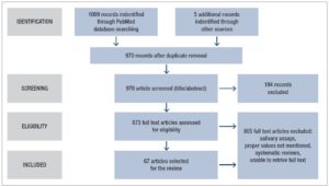
Articles identified from the keyword search were transferred to an Endnote database. Once duplicates were removed, the title, keywords and abstracts were reviewed for suitability for inclusion. All full texts except for 18 were accessed and reviewed for suitability (considering gender and age group of samples, and reporting of T levels). Forward citations of the studies retrieved were also traced and screened for possible inclusion and five additional articles were identified for inclusion.  The search was conducted independently by KA and LA and the articles to be included in the review were determined through an interactive consensus process involving KA, NS, LA and DMSF (Fig.1).


Inclusion and exclusion criteria

For ease of comparability, the review included only research articles published in English and referring to serum samples, even though sputum or salivary T levels are considered good surrogates for serum T. Conference proceedings, editorials, commentaries, book chapters and book reviews were excluded. Studies describing two or more serum T readings across the MC were included even though such measurement was not the main objective of those selected studies. Studies that did not mention T values separately for each phase of the MC were excluded.


Data extraction

The full texts of the selected articles were reviewed and relevant variables were extracted into a datasheet. Extracted variables included, author/s, year of publication, method of detection of serum T, sample size, demographics of the sample, sample collection method, MC phase in which the sample was collected, and T values recorded.

Results

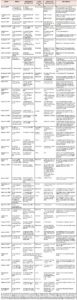
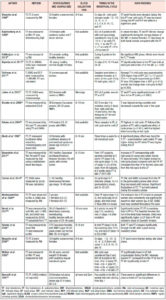
The primary search identified 1009 articles which were reviewed for duplicates, after which the title, keywords and abstracts of the remaining articles were reviewed. Five additional articles were identified by manually searching the reference lists and forward citations of included papers. A total of 67 articles were included in the review. Tables 1 and 2 summarize the studies included in the review.


Characteristics of studies

The included studies were conducted between years 1972 and 2020 and the number of participants ranged from 6 to 259. The studies concerned different ethnicities from North and South America, Europe, the Middle East, Asia and Australia. The females included were aged 18-45 years with a BMI ranging from 18 to 35 kg/m2. The T assessment methods varied between studies.


Timing in the menstrual cycle

Testosterone measurements were done at variable time points in the MC and few studies reported daily or every other day (EOD) serum sampling[36-39]. Some studies assessed T at four points in the MC i.e., early and late in the follicular phase (FP) and early and late in the luteal phase (LP) [40-42]; some at three points, i.e., early FP , mid cycle (close to ovulation) and LP [43-45]; and others at two points, i.e., in middle of the FP and the LP respectively [46-48].

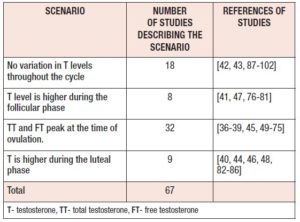
Multiple studies report that serum TT and FT levels peak during mid cycle at ovulation [36-39, 45, 49-75]. However, T level variation during the rest of the cycle does not seem to show a consistent pattern.  Eight studies reported higher values during the FP[41, 47, 76-81], while nine studies reported higher values during the LP[40, 44, 46, 48, 82-86]. (Table 3). Many studies reported no change in T across the MC[42, 43, 87-102]. Out of the eighteen studies which did not observe any cyclical change, eight had assayed only two points (namely FP and LP) [89, 90, 92-95, 97, 98] and only four studies had sampled daily or EOD [87, 88, 96, 97].


Serum testosterone assessment technique

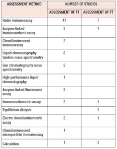
The included studies reported multiple methods of detecting serum T (Table 4). Studies which compared T detection methods, used more than one measurement method for T detection. Few studies assessed both TT and FT [37, 41, 42, 44, 47, 57, 68, 71, 78, 89], while the majority tested only TT [40, 43, 53, 61, 65, 70, 76, 79, 80, 83, 86, 91, 93].


Discussion

Testosterone peak at mid cycle

Most studies reported at least a minute mid-cycle increase in TT, which coincided with ovulation. Irrespective of the sample size and detection method, TT peaked during mid cycle in the studies where daily sampling was done. Studies which sampled T at two points during the MC did not observe a T peak as this could not be demonstrated by plotting only two measurements. Levels of T were lower in late LP and in the peri menstrual phase [61] and lowest close to menstruation consistently. Daily sampling is more accurate as the increased number of samples overcomes the diurnal variation and sampling bias.

The mid-cycle T increase is due to synthesis of T by the ovarian theca cells, mainly regulated by LH [10]. LH receptors are located on the theca cells during all stages of the MC. LH principally stimulates androstenedione production and, to a lesser degree, production of T in theca cells. Androstenedione is then transported to the granulosa cells where it is aromatized to estrone and then converted to estradiol by 17-β-hydroxysteroid dehydrogenase. In contrast, in the granulosa cells LH receptors only appear in mature follicles greater than 10 mm in diameter, i.e., in the antral follicle, which is most likely to develop as the dominant follicle[41, 103]. Ovarian androgen production and its conversion to estradiol are essential for follicle recruitment and the process of ovulation. Insufficient androgen production in the FP [14] and high androgen levels can lead to anovulation[104, 105]. Testosterone was significantly higher during the FP in females with anovulatory menstrual cycles [55].

Ovulation is triggered by the LH surge and it is plausible that a LH-mediated increase in ovarian androgen production occurs. The peak in T is unlikely to be due to adrenal androgen production or peripheral aromatization [69]. The mid-cycle androgen rise may accelerate follicular atresia and ensure that a single dominant follicle reaches the point of ovulation[68]. It is likely that androgens facilitate apoptosis and consequent follicle atresia only in mature antral follicles [104]. The mid-cycle increase in sexual activity could be attributed to the mid-cycle androgen spike[106, 107]. If high T is related to the LH surge at ovulation it must gradually recede from the FP to the LP and reported measurements showed both TT and FT to follow this pattern until the next FP [50, 60]. A reduction in SHBG is also reported in the mid cycle, coinciding with ovulation [45, 50] and possibly further increasing the FT levels, followed by an increase in SHBG levels during the LP [99].

The T rise in the mid cycle could affect smooth muscle and may assist ovulation and subsequent actions on the fallopian tube. The mechanism of action of T on smooth muscle and the inflammatory cascade needs to be studied further to understand the physiological basis for the rise in T during the MC.

The mid-cycle rise in FT was more prominent in younger females while it was not evident in older females aged between 43 and 47 years, who were cycling regularly and had normal levels of prolactin and thyroid-stimulating hormone [68]. The decreased concentrations of FT and androstenedione, without significant changes in SHBG, suggest that in older women these hormones are produced in lower quantities during the mid-cycle. It was concluded that this process was ovary dependent and not adrenal dependent as the observed changes varied with the stage of the MC.


Variation in reported mid-cycle testosterone values

Mid-cycle FT has been reported to be 63ng/dL (23-120ng/dL) [50] and as low as 23.72 ± 15.35 ng/dL [52]. However, most of the values reported in the included studies were within the currently accepted reference ranges (0.5-2.5 nmol/L or 15-75 ng/dL) [20]. The variability of reported serum FT values was dependent on several factors such as study design, study sample (age and BMI), assay techniques and their limitations, low T threshold and its statistical interpretation. The studies incorporated in the review mainly concerned apparently healthy females. Obesity, hypothyroidism, acromegaly, and diabetes mellitus, as well as glucocorticoids and anabolic steroids are known to decrease SHBG, resulting in low total T and high FT levels; SHBG levels are also known to decrease with old age, liver disease, and anticonvulsant and estrogen use, resulting in altered T profiles [18]. Higher levels of TT and FT with low SHBG have been reported in females with polycystic ovarian syndrome (PCOS) [108]. Studies involving women with chronic medical conditions, metabolic comorbidities like PCOS, diabetes mellitus, and other endocrine abnormalities are not within the scope of this review, and were therefore not included. However, BMI was seen to vary with different studies, with some studies also including females with higher BMI values. Of the limited number of studies which also evaluated overweight and obese females, some studies reported low TT [86], while others reported consistently high TT and FT values throughout the MC [41].


Variation of SHBG

An increase in SHBG in the LP compared with the FP was reported for ovulatory MCs [47, 54, 98, 109]. However, no consistent pattern of changes in SHBG, TT or FT levels was observed. Only a few studies had a sample size of more than 20 [42, 44-51, 77, 83], which could also affect these reported outcomes.

Timing of collection of samples during the menstrual cycle

As regards the definitions of the phases of the MC and the timing of sample collections, the studies showed considerable differences. When the FP was considered to last from day 0 to ovulation and the LP from ovulation to beginning of next menstrual cycle, there was uncertainty over the best day to sample in each of these two phases. Some studies, however, considered a range of days after the beginning of the MC for drawing blood in the FP, while mid cycle was defined as day 14-15 and the LP as after the mid cycle. The mid cycle was considered the late FP in some studies. In most studies at least three samples were taken, one during the FP, one at mid cycle, and one during the LP [43-45]. Instead, the few studies analyzing daily blood samples did not encounter the problem of determining ovulation and provided the best evidence for changing patterns of T [39, 62, 63, 100].


The selection of females

Most studies included females of reproductive age with regular menstrual cycles and they ranged from adolescents to premenopausal females. The participants also varied in terms of marital status and parity.

Although changes in T depend on whether a cycle is ovulatory or anovulatory, most of the studies did not identify MCs as ovulatory or anovulatory. Studies that did identify ovulatory and anovulatory cycles reported different patterns of T fluctuation, where in anovulatory cycles no mid-cycle rise in T was seen, with one study reporting higher T during the FP [56]. In contrast, in some studies no difference in T were seen between the females with ovulatory and anovulatory cycles [67].

BMI, smoking, alcohol consumption and exercise are known confounders for T levels in females [24]. However, very few studies have controlled for such confounding factors. Non-smoking females, without any chronic medical disease, who were not on any medication were studied in well planned studies omitting the errors due to the above factors [52, 78].

Sample timing during the day

It is important that timing of the sampling is uniform at all points of the MC as hormone levels are known to decrease after waking due to diurnal variation [110, 111]. In most studies sampling was done in the morning to get the highest T levels, and after an overnight fast. Fasting overnight was important as food interferes with the results of the analysis [112]. Some studies did sampling throughout the day and these provide the best evidence for the highest peak of T during the day.


Method of assessing testosterone

Most studies used the RIA method to measure TT, while some used the gold standard LC-MS/MS. Others used ELISA, CLIA, HPLC, ELFA. Older studies used RIA with organic extraction and chromatographic separation to remove interfering substances and matrix effects.[20] RIA has sensitivity to detect higher concentrations of hormone while errors may occur at lower concentrations especially in the late LP when hormone concentrations are lower. Recent studies have used automated platforms with nonradioactive methods like chemiluminescent detection. Liquid chromatography mass spectrometry (LC-MS) and  LC-MS/MS can assess low concentrations  of T  seen in the MC [20].

It was difficult to compare studies as the assay techniques used and the T fraction tested (TT or FT) differed between studies; furthermore, outcomes were expressed in different forms, such as the T ratio or the FT index (calculated from TT and SHBG).

Future research on T changes during the MC needs to be designed to address the current knowledge gaps. MC phases and timing of blood sampling should be well defined. Future study protocols should include at least three sampling points per MC as a peak cannot be demonstrated when plotting only two measurements. Daily sampling, however, will overcome problems encountered in defining the phases of the MC and in detecting ovulation for mid-cycle sampling. The use of daily, morning samples, taken from healthy regularly menstruating females and tested using the liquid chromatography method, is recommended as the best option to achieve coherent results.


Conclusions

Both TT and FT peak at ovulation in females with regular (ovulatory) menstrual cycles. In the early LP, TT levels appear to be higher than in the early FP, but levels during the late LP seem to be lower than in the mid-follicular phase. Variations in serum T levels during different phases of the menstrual cycle seem to be related to whether the MC is ovulatory or anovulatory, to the T assessment technique used, and the timing of sample collection both during the MC and during the day. Further research should be conducted on larger populations and using more clearly defined criteria.

Conflict of interest

The authors declare that there is no conflict of interest.

Media

References

  1. Burger HG. Androgen production in women. Fertil Steril. 2002;77 Suppl 4:S3-5.
  2. Sowers MF, Beebe JL, McConnell D, Randolph J, Jannausch M. Testosterone concentrations in women aged 25-50 years: associations with lifestyle, body composition, and ovarian status. Am J Epidemiol. 2001;153:256-64.
  3. Hammes SR, Levin ER. Impact of estrogens in males and androgens in females. J Clin Invest. 2019;129:1818-26.
  4. Glaser R, Dimitrakakis C. Testosterone therapy in women: myths and misconceptions. Maturitas. 2013;74:230-4.
  5. Hodgson KT, Braunstein GD. Physiological effects of androgens in women. In: Azziz R, Nestler JE, Dewailly D, eds. Androgen excess disorders in women. Contemporary endocrinology. Humana Press; 2006:49-62.
  6. Jeffcoate SL BR, Lim NY, London DR, Prunty ITO, Spathis OS. Androgen production in hypogonadal men. J Endocrinol. 1967;37:401-11.
  7. Brooks R. Androgens: physiology and pathology. In: Makin HLJ, ed. Biochemistry of steroid hormones Blackwell Scientific, Oxford; 1984:565-94.
  8. Wheeler MJ. The determination of bio-available testosterone. Ann Clin Biochem. 1995;32:345-57.
  9. Ducharme JR, Forest MG, De Peretti E, Sempé M, Collu R, Bertrand J. Plasma adrenal and gonadal sex steroids in human pubertal development. J Clin Endocrinol Metab. 1976;42:468-76.
  10. Schiebinger RJ, Albertson BD, Cassorla FG, et al. The developmental changes in plasma adrenal androgens during infancy and adrenarche are associated with changing activities of adrenal microsomal 17-hydroxylase and 17-20 desmolase. J Clin Invest. 1981;67:1177-82.
  11. Angelopoulos N, Goula A, Tolis G. The role of luteinizing hormone activity in controlled ovarian stimulation. J Endocrinol Invest. 2005;28:79-88.
  12. Tan S, Hahn S, Janssen OE. Insulin resistance syndrome and polycystic ovary syndrome: implications for diagnosis and treatment. Panminerva Med. 2005;47:211-7.
  13. McNatty KP, Makris A, DeGrazia C, Osathanondh R, Ryan KJ. The production of progesterone, androgens, and estrogens by granulosa cells, thecal tissue, and stromal tissue from human ovaries in vitro. J Clin Endocrinol Metab. 1979;49:687-99.
  14. Haning RV Jr, Hackett RJ, Flood CA, Loughlin JS, Zhao QY, Longcope C. Testosterone, a follicular regulator: key to anovulation. J Clin Endocrinol Metab. 1993;77:710-5.
  15. Vermeulen A. Evaluation of plasma testosterone: which test and when? Current Sexual Health Reports. 2005;2:37-40.
  16. Montanini V, Simoni M, Chiossi G, et al. Age-related changes in plasma dehydroepiandrosterone sulphate, cortisol, testosterone and free testosterone circadian rhythm in adult men. Horm Res. 1988;29:1-6.
  17. Bodenheimer S WJ, Faiman C. Diurnal rhythms of serum gonadotrophins and testosterone, estradiol and cortisol in blind men. J Clin Endocrinol Metab. 1973;37:472-5.
  18. Krakowsky Y, Grober ED. Testosterone deficiency – establishing a biochemical diagnosis. EJIFCC. 2015;26:105-13.
  19. Kanakis GA, Tsametis CP, Goulis DG. Measuring testosterone in women and men. Maturitas. 2019;125:41-4.
  20. Matsumoto AM, Bremner WJ. Serum testosterone assays–accuracy matters. J Clin Endocrinol Metab. 2004;89:520-4.
  21. Rouleau M, Lemire F, Déry M, et al. Discordance between testosterone measurement methods in castrated prostate cancer patients. Endocr Connect. 2019;8:132-40.
  22. Wang C, Catlin DH, Demers LM, Starcevic B, Swerdloff RS. Measurement of total serum testosterone in adult men: comparison of current laboratory methods versus liquid chromatography-tandem mass spectrometry. J Clin Endocrinol Metab. 2004;89:534-43.
  23. Cupisti S, Dittrich R, Binder H, et al. Influence of body mass index on measured and calculated androgen parameters in adult women with Hirsutism and PCOS. Exp Clin Endocrinol Diabetes. 2007;115:380-6.
  24. Trost LW, Mulhall JP. Challenges in testosterone measurement, data interpretation, and methodological appraisal of interventional trials. J Sex Med. 2016;13:1029-46.
  25. Canguven O, Albayrak S. Do low testosterone levels contribute to the pathogenesis of asthma? Med Hypotheses. 2011;76:585-8.
  26. Ono YJ, Tanabe A, Nakamura Y, et al. A low-testosterone state associated with endometrioma leads to the apoptosis of granulosa cells. PLoS One. 2014;9:e115618.
  27. Imrich R, Rovensky J, Malis F, et al. Low levels of dehydroepiandrosterone sulphate in plasma, and reduced sympathoadrenal response to hypoglycaemia in premenopausal women with rheumatoid arthritis. Ann Rheum Dis. 2005;64:202-6.
  28. Lahita RG, Bradlow HL, Ginzler E, Pang S, New M. Low plasma androgens in women with systemic lupus erythematosus. Arthritis Rheum. 1987;30:241-8.
  29. Masi AT, Chatterton RT, Aldag JC. Perturbations of hypothalamic-pituitary-gonadal axis and adrenal androgen functions in rheumatoid arthritis: an odyssey of hormonal relationships to the disease. Ann N Y Acad Sci. 1999;876:53-62; discussion 62-3.
  30. Spector TD, Perry LA, Tubb G, Silman AJ, Huskisson EC. Low free testosterone levels in rheumatoid arthritis. Ann Rheum Dis. 1988;47:65-8.
  31. Tomassini V, Onesti E, Mainero C, et al. Sex hormones modulate brain damage in multiple sclerosis: MRI evidence. J Neurol Neurosurg Psychiatry. 2005;76:272-5.
  32. Perković D, Martinović Kaliterna D, Jurišić Z, Lalovac M, Radić M. Androgens in post-menopausal patients with systemic sclerosis. Rheumatology (Oxford). 2015;54:744-6.
  33. Rasmussen JJ, Selmer C, Frøssing S, et al. Endogenous testosterone levels are associated with risk of type 2 diabetes in women without established comorbidity. J Endocr Soc. 2020;4:bvaa050.
  34. Armstrong PW, Ezekowitz JA. Testosterone therapy in women with heart failure “why can’t a woman be more like a man?”. J Am Coll Cardiol. 2010;56:1317-9.
  35. Bianchi VE. The anti-inflammatory effects of testosterone. J Endocr Soc. 2018;3:91-107.
  36. Bui HN, Sluss PM, Blincko S, Knol DL, Blankenstein MA, Heijboer AC. Dynamics of serum testosterone during the menstrual cycle evaluated by daily measurements with an ID-LC-MS/MS method and a 2nd generation automated immunoassay. Steroids. 2013;78:96-101.
  37. Massafra C, De Felice C, Agnusdei DP, Gioia D, Bagnoli F. Androgens and osteocalcin during the menstrual cycle. J Clin Endocrinol Metab. 1999;84:971-4.
  38. Goebelsmann U, Arce JJ, Thorneycroft IH, Mishell DR Jr. Serum testosterone concentrations in women throughout the menstrual cycle and following HCG administration. Am J Obstet Gynecol. 1974;119:445-52.
  39. Judd HL, Yen SS. Serum androstenedione and testosterone levels during the menstrual cycle. J Clin Endocrinol Metab. 1973;36:475-81.
  40. Oka K, Hirano T, Noguchi M. Changes in the concentration of testosterone in serum during the menstrual cycle, as determined by liquid chromatography. Clin Chem. 1988;34:557-60.
  41. Wajchenberg BL, Marcondes JA, Mathor MB, Achando SS, Germak OA, Kirschner MA. Free testosterone levels during the menstrual cycle in obese versus normal women. Fertil Steril. 1989;51:535-7.
  42. Veldhuijzen DS, Keaser ML, Traub DS, Zhuo J, Gullapalli RP, Greenspan JD. The role of circulating sex hormones in menstrual cycle-dependent modulation of pain-related brain activation. Pain. 2013;154:548-59.
  43. Nóbrega LH, Azevedo GD, Lima JG, et al. Analysis of testosterone pulsatility in women with ovulatory menstrual cycles. Arq Bras Endocrinol Metabol. 2009;53:1040-6.
  44. Wyskida K, Franik G, Wikarek T, et al. The levels of adipokines in relation to hormonal changes during the menstrual cycle in young, normal-weight women. Endocr Connect. 2017;6:892-900.
  45. Rothman MS, Carlson NE, Xu M, et al. Reexamination of testosterone, dihydrotestosterone, estradiol and estrone levels across the menstrual cycle and in postmenopausal women measured by liquid chromatography-tandem mass spectrometry. Steroids. 2011;76:177-82.
  46. Matteis M, Polverino F, Spaziano G, et al. Effects of sex hormones on bronchial reactivity during the menstrual cycle. BMC Pulm Med. 2014;14:108.
  47. Linton L, Taylor M, Dunn S, et al. Associations of serum levels of sex hormones in follicular and luteal phases of the menstrual cycle with breast tissue characteristics in young women. PLoS One. 2016;11:e0163865.
  48. Edam KA, Kredy HM, Alhamami MM. Clinical study for testosterone, progesterone and oxidative stress during phases of menstrual cycle in women with bronchial asthma. Indian Journal of Public Health Research & Development. 2019;10:3047-52.
  49. Šrámková M, Dušková M, Vítků J, et al. Levels of adipokines and some steroids during the menstrual cycle. Physiol Res. 2015;64:S147-54.
  50. Wunder DM, Yared M, Bersinger NA, Widmer D, Kretschmer R, Birkhäuser MH. Serum leptin and C-reactive protein levels in the physiological spontaneous menstrual cycle in reproductive age women. Eur J Endocrinol. 2006;155:137-42.
  51. Shultz SJ, Gansneder BM, Sander TC, Kirk SE, Perrin DH. Absolute serum hormone levels predict the magnitude of change in anterior knee laxity across the menstrual cycle. J Orthop Res. 2006;24:124-31.
  52. Muneyvirci-Delale O, Nacharaju VL, Altura BM, Altura BT. Sex steroid hormones modulate serum ionized magnesium and calcium levels throughout the menstrual cycle in women. Fertil Steril. 1998;69:958-62.
  53. Ahrens KA, Vladutiu CJ, Mumford SL, et al. The effect of physical activity across the menstrual cycle on reproductive function. Ann Epidemiol. 2014;24:127-34.
  54. Adams JM, Taylor AE, Crowley WF Jr, Hall JE. Polycystic ovarian morphology with regular ovulatory cycles: insights into the pathophysiology of polycystic ovarian syndrome. J Clin Endocrinol Metab. 2004;89:4343-50.
  55. Apter D, Viinikka L, Vihko R. Hormonal pattern of adolescent menstrual cycles. J Clin Endocrinol Metab. 1978;47:944-54.
  56. Bancroft J, Sanders D, Davidson D, Warner P. Mood, sexuality, hormones, and the menstrual cycle. III. Sexuality and the role of androgens. Psychosom Med. 1983;45:509-16.
  57. Bloch M, Schmidt PJ, Su TP, Tobin MB, Rubinow DR. Pituitary-adrenal hormones and testosterone across the menstrual cycle in women with premenstrual syndrome and controls. Biol Psychiatry. 1998;43:897-903.
  58. Braunstein GD, Reitz RE, Buch A, Schnell D, Caulfield MP. Testosterone reference ranges in normally cycling healthy premenopausal women. J Sex Med. 2011;8:2924-34.
  59. Brideau NA, Forest JC, Lemay A, Dodin S. Correlation between ovarian steroids and lipid fractions in relation to age in premenopausal women. Clin Endocrinol (Oxf). 1992;37:437-44.
  60. Caruso S, Agnello C, Malandrino C, Lo Presti L, Cicero C, Cianci S. Do hormones influence women’s sex? Sexual activity over the menstrual cycle. J Sex Med. 2014;11:211-21.
  61. Dougherty DM, Bjork JM, Moeller FG, Swann AC. The influence of menstrual-cycle phase on the relationship between testosterone and aggression. Physiol Behav. 1997;62:431-5.
  62. Epstein MT, McNeilly AS, Murray MA, Hockaday TD. Plasma testosterone and prolactin in the menstrual cycle. Clin Endocrinol (Oxf). 1975;4:531-5.
  63. Frölich M, Brand EC, van Hall EV. Serum levels of unconjugated aetiocholanolone androstenedione, testosterone, dehydroepiandrosterone, aldosterone, progesterone and oestrogens during the normal menstrual cycle. Acta Endocrinol (Copenh). 1976;81:548-62.
  64. Genazzani AR, Inaudi P, D’Ambrogio G, Romagnino S, Nasi A, Facchinetti F. Plasma androgens and menstrual cycle: physiopathological correlates from adolescence to menopause. Horm Res. 1983;18:84-97.
  65. Guapo VG, Graeff FG, Zani AC, Labate CM, dos Reis RM, Del-Ben CM. Effects of sex hormonal levels and phases of the menstrual cycle in the processing of emotional faces. Psychoneuroendocrinology. 2009;34:1087-94.
  66. Kim MH, Rosenfield RL, Dupon C. The effects of dexamethasone on plasma free androgens during the normal menstrual cycle. Am J Obstet Gynecol. 1976;126:982-6.
  67. Kurzer MS, Calloway DH. Effects of energy deprivation on sex hormone patterns in healthy menstruating women. Am J Physiol. 1986;251:E483-8.
  68. Mushayandebvu T, Castracane VD, Gimpel T, Adel T, Santoro N. Evidence for diminished midcycle ovarian androgen production in older reproductive aged women. Fertil Steril. 1996;65:721-3.
  69. Rezaii T, Gustafsson TP, Axelson M, et al. Circulating androgens and SHBG during the normal menstrual cycle in two ethnic populations. Scand J Clin Lab Invest. 2017;77:184-9.
  70. Rubinow DR, Hoban MC, Grover GN, et al. Changes in plasma hormones across the menstrual cycle in patients with menstrually related mood disorder and in control subjects. Am J Obstet Gynecol. 1988;158:5-11.
  71. Salonia A, Pontillo M, Nappi RE, et al. Menstrual cycle-related changes in circulating androgens in healthy women with self-reported normal sexual function. J Sex Med. 2008;5:854-63.
  72. Schreiner-Engel P, Schiavi RC, Smith H, White D. Sexual arousability and the menstrual cycle. Psychosom Med. 1981;43:199-214.
  73. Schreiner-Engel P, Schiavi RC, White D, Ghizzani A. Low sexual desire in women: the role of reproductive hormones. Horm Behav. 1989;23:221-34.
  74. Van Goozen SH, Wiegant VM, Endert E, Helmond FA, Van de Poll NE. Psychoendocrinological assessment of the menstrual cycle: the relationship between hormones, sexuality, and mood. Arch Sex Behav. 1997;26:359-82.
  75. Vermeulen A, Verdonck L. Plasma androgen levels during the menstrual cycle. Am J Obstet Gynecol. 1976;125:491-4.
  76. Dušková M, Simůnková K, Hill M, et al. Chronic cigarette smoking alters circulating sex hormones and neuroactive steroids in premenopausal women. Physiol Res. 2012;61:97-111.
  77. Okifuji A, Turk DC. Sex hormones and pain in regularly menstruating women with fibromyalgia syndrome. J Pain. 2006;7:851-9.
  78. Massafra C, Gioia D, De Felice C, et al. Effects of estrogens and androgens on erythrocyte antioxidant superoxide dismutase, catalase and glutathione peroxidase activities during the menstrual cycle. J Endocrinol. 2000;167:447-52.
  79. Hill M, Havlíková H, Vrbíková J, et al. The identification and simultaneous quantification of 7-hydroxylated metabolites of pregnenolone, dehydroepiandrosterone, 3beta,17beta-androstenediol, and testosterone in human serum using gas chromatography-mass spectrometry. J Steroid Biochem Mol Biol. 2005;96:187-200.
  80. Kannenberg F, Fobker M, Schulte E, et al. The Simultaneous measurement of serum testosterone and 5α-dihydrotestosterone by gas chromatography-mass spectrometry (GC-MS). Clin Chim Acta. 2018;476:15-24.
  81. Mathor MB AS, Wajchenberg BL, Germek OA. Free plasma testosterone levels during the normal menstrual cycle. Journal of Endocrinological Investigation. 1985;8:437-41.
  82. Hietala M, Sandberg T, Borg A, Olsson H, Jernström H. Testosterone levels in relation to oral contraceptive use and the androgen receptor CAG and GGC length polymorphisms in healthy young women. Hum Reprod. 2007;22:83-91.
  83. Salonia A, Lanzi R, Scavini M, et al. Sexual function and endocrine profile in fertile women with type 1 diabetes. Diabetes Care. 2006;29:312-6.
  84. Anttila L, Koskinen P, Irjala K, Kaihola HL. Reference intervals for serum sex steroids and gonadotropins in regularly menstruating women. Acta Obstet Gynecol Scand. 1991;70:475-81.
  85. Apter D, Jánne O, Karvonen P, Vihko R. Simultaneous determination of five sex hormones in human serum by radioimmunoassay after chromatography on Lipidex-5000. Clin Chem. 1976;22:32-8.
  86. Skiba MA, Bell RJ, Islam RM, Handelsman DJ, Desai R, Davis SR. Androgens during the reproductive years: what is normal for women? J Clin Endocrinol Metab. 2019;104:5382-92.
  87. Fuller-Jackson JP, Dordevic AL, Clarke IJ, Henry BA. Effect of sex and sex steroids on brown adipose tissue heat production in humans. Eur J Endocrinol. 2020;183:343-55.
  88. Arnoni-Bauer Y, Bick A, Raz N, et al. Is it me or my hormones? Neuroendocrine activation profiles to visual food stimuli across the menstrual cycle. J Clin Endocrinol Metab. 2017;102:3406-14.
  89. Bancroft J, Sherwin BB, Alexander GM, Davidson DW, Walker A. Oral contraceptives, androgens, and the sexuality of young women: II. The role of androgens. Arch Sex Behav. 1991;20:121-35.
  90. Collins A, Eneroth P, Landgren BM. Psychoneuroendocrine stress responses and mood as related to the menstrual cycle. Psychosom Med. 1985;47:512-27.
  91. Dada OA, Osinusi BO, Nduka EU, Osotimehin BO, Ladipo OA. 17 Beta-estradiol, progesterone and testosterone in the normal menstrual cycle of Nigerians. Int J Gynaecol Obstet. 1984;22:151-4.
  92. Dawood MY, Saxena BB. Plasma testosterone and dihydrotestosterone in ovulatory and anovulatory cycles. Am J Obstet Gynecol. 1976;126:430-5.
  93. Elliott KJ, Cable NT, Reilly T, Diver MJ. Effect of menstrual cycle phase on the concentration of bioavailable 17-beta oestradiol and testosterone and muscle strength. Clin Sci (Lond). 2003;105:663-9.
  94. Fahmy K, Ghoneim M, Eisa I, el-Gazar A, Afifi A. Serum and endometrial sodium and potassium levels with inert and copper-containing IUCDs and relation to serum steroid levels. Contraception. 1992;45:573-81.
  95. Feldman F, Bain J, Matuk AR. Daily assessment of ocular and hormonal variables throughout the menstrual cycle. Arch Ophthalmol. 1978;96:1835-8.
  96. Luthold WW, Borges MF, Marcondes JA, Hakohyama M, Wajchenberg BL, Kirschner MA. Serum testosterone fractions in women: normal and abnormal clinical states. Metabolism. 1993;42:638-43.
  97. Phillips SM, Sherwin BB. Variations in memory function and sex steroid hormones across the menstrual cycle. Psychoneuroendocrinology. 1992;17:497-506.
  98. Schijf CP, van der Mooren MJ, Doesburg WH, Thomas CM, Rolland R. Differences in serum lipids, lipoproteins, sex hormone binding globulin and testosterone between the follicular and the luteal phase of the menstrual cycle. Acta Endocrinol (Copenh). 1993;129:130-3.
  99. Schöning S, Engelien A, Kugel H, et al. Functional anatomy of visuo-spatial working memory during mental rotation is influenced by sex, menstrual cycle, and sex steroid hormones. Neuropsychologia. 2007;45:3203-14.
  100. Tyler JP, Newton JR, Collins WP. Variations in the concentration of testosterone in peripheral venous plasma from healthy women. Acta Endocrinol (Copenh). 1975;80:542-50.
  101. Valette A, Seradour B, Boyer J. Plasma testosterone levels during the menstrual cycle. J Clin Endocrinol Metab. 1975;40:160-1.
  102. Werawatgoompa S, Tankeyoon M, Kongseripong R, et al. Hormonal changes in normal menstrual cycle of Thai women. Contraception. 1981;23:301-13.
  103. Franks S, Hardy K. Androgen action in the ovary. Front Endocrinol (Lausanne). 2018;9:452.
  104. Gleicher N, Weghofer A, Barad DH. The role of androgens in follicle maturation and ovulation induction: friend or foe of infertility treatment? Reprod Biol Endocrinol. 2011;9:116.
  105. Rosenfield RL, Ehrmann DA. The pathogenesis of Polycystic Ovary Syndrome (PCOS): the hypothesis of PCOS as functional ovarian hyperandrogenism revisited. Endocr Rev. 2016;37:467-520.
  106. Adams DB, Gold AR, Burt AD. Rise in female-initiated sexual activity at ovulation and its suppression by oral contraceptives. N Engl J Med. 1978;299:1145-50.
  107. Morris NM, Udry JR, Khan-Dawood F, Dawood MY. Marital sex frequency and midcycle female testosterone. Arch Sex Behav. 1987;16:27-37.
  108. Penttilä TA, Anttila L, Törmä A, Koskinen P, Erkkola R, Irjala K. Serum free testosterone in polycystic ovary syndrome measured with a new reference method. Fertil Steril. 1996;65:55-60.
  109. Plymate SR, Moore DE, Cheng CY, Bardin CW, Southworth MB, Levinski MJ. Sex hormone-binding globulin changes during the menstrual cycle. J Clin Endocrinol Metab. 1985;61:993-6.
  110. Panico S, Pisani P, Muti P, et al. Diurnal variation of testosterone and estradiol: a source of bias in comparative studies on breast cancer. J Endocrinol Invest. 1990;13:423-6.
  111. Brambilla DJ, Matsumoto AM, Araujo AB, McKinlay JB. The effect of diurnal variation on clinical measurement of serum testosterone and other sex hormone levels in men. J Clin Endocrinol Metab. 2009;94:907-13.
  112. Lehtihet M, Arver S, Bartuseviciene I, Pousette A. S-testosterone decrease after a mixed meal in healthy men independent of SHBG and gonadotrophin levels. Andrologia. 2012;44:405-10.

Keywords: , , ,

Citation: R Atukorala K.,Silva W.,Amarasiri L., D., Changes in serum testosterone during the menstrual cycle – an integrative systematic review of published literature, GREM Gynecological and Reproductive Endocrinology & Metabolism (2022); 01/2022:009-020 doi: 10.53260/grem.223012

Published: March 14, 2022