Review, 010-014 | DOI: 10.53260/grem.2450102


Role of mitochondria and redox state in sperm and oocyte health

Abstract

Mitochondria are the “powerhouse” of eukaryote cells and, according to the endosymbiont hypothesis, developed from aerobic bacteria or incorporated factual anaerobic bacteria. Mitochondria do not only produce energy in form of ATP, but also a small amount of reactive oxygen species (ROS) that normally trigger essential physiological reactions. In case, of excessive ROS production or too low availability of antioxidants, this will lead to oxidative stress which causes damage to all biomolecules including nuclear and mitochondrial DNA. As a result, mitochondrial ROS production is further increased, and redox homeostasis moves toward oxidative stress. This condition can be caused by a number of health (e.g. polycystic ovary syndrome [PCOS], endometriosis) and poor lifestyle (e.g. obesity, smoking) conditions and has serious consequences for the male and female reproductive system. In sperm, a small amount of ROS is normally triggering functions such as capacitation and acrosome reaction whereas oxidative stress is causing sperm DNA fragmentation. Similarly, in the ovaries, a limited amount of ROS is necessary for the regulation of the ovarian cycle and steroidogenesis, while a higher amount of ROS is involved in ovarian aging, and cause mitochondrial dysfunction, spindle abnormalities or telomere shortening. L-Carnitine as an antioxidant has been shown to significantly improve male and female reproductive functions as this quaternary amine not only has direct antioxidant properties but is also closely involved in shuttling long-chain fatty acids into the mitochondrial β-oxidation for energy production as well as the detoxification of damaging lipids.

Full article PDF

Full Article

Introduction

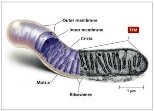
In all eukaryote cells, mitochondria are the organelles providing most of the energy used by these cells and are therefore also called the “powerhouse of the cells”. In terms of evolutionary biology, these organelles originate either from endosymbiotically living aerobic bacteria or from incorporated factual anaerobic bacteria in archaea [1]. Structurally, mitochondria have two membranes, an outer membrane and an inner membrane forming so-called cristae which are highly compartmentalized sections (Figure 1). Between these two membranes is the intermembrane space. The space enclosed by the inner membrane is called matrix. Human sperm contain about 50 to 75 mitochondria helically arranged around the axoneme in their midpiece in about 12 to 13 gyres and 2 mitochondria per gyre [2,3]. In contrast, mature human oocytes contain about 100,000 spherical mitochondria with only a small number of cristae [4].

In the cytoplasm of cells, glycolysis is an anaerobic process taking place converting glucose into pyruvate and producing four molecules of adenosine triphosphate (ATP). In the presence of oxygen, the α-keto acid pyruvate is transported by pyruvate carriers into the mitochondrial matrix and then converted by pyruvate dehydrogenase into acetyl-CoA which enters the Krebs cycle for oxidative phosphorylation (OXPHOS) [6]. Acetyl-CoA is also formed via β-oxidation from fatty acids which are transported through the inner mitochondrial membrane by carnitine/acylcarnitine [7]. The multi-enzyme complexes I-IV (complex I: NADH dehydrogenase; complex II: succinate dehydrogenase; complex III: cytochrome 3 reductase; complex IV: cytochrome 3 oxidase) located in the inner mitochondrial membrane are forming the mitochondrial electron transport chain (ETC), while complex V (ATP synthase) produces the ATP that is necessary for cellular energy needs [8].

As a result of this oxidative phosphorylation, not only ATP is produced but up to 5% of the consumed oxygen is leaving the system as superoxide (•O2-), hydrogen peroxide (H2O2) or other reactive oxygen species (ROS) compounds [9-11], which have half-life times in the milli-second (10-3 s) to nano-second (10-9 s) range [12]. These cytotoxic products accelerate cell aging and can cause numerous disorders and cell death via cell cycle dysregulation and apoptosis [13]. Therefore, with the significant increase in the atmospheric oxygen concentration about 2.3 billion years ago [14], protective mechanisms preventing extremely sensitive biomolecules developed including enzymatic and non-enzymatic antioxidant systems. Life also adapted to these higher oxygen levels by using it to produce energy in the mitochondria [15] as well as for cellular redox regulation, signaling [16] and triggering essential physiological functions such as gene regulation, cellular activities or synaptic plasticity [17-20]. Hence, H2O2 and •O2- are not only harmful, but fulfill essential physiological and cellular functions regulating functions such as immunity, proliferation, development, or steroidogenesis [21]; all depending on the concentration.

Since mitochondria developed from endosymbiotic bacteria, mitochondria have their own DNA (mtDNA) which encodes for approximately 13 protein subunits of the mitochondrial electron transfer chain, and ribosomal RNA (rRNA) and transfer RNA (tRNA) components of the mitochondrial translation system [22]. However, contrary to nuclear DNA (nDNA), mtDNA has 3 to 7 circular, much shorter, double-stranded DNA strands which are and not protected by histones and protamines like the nDNA. Furthermore, since mtDNA replicates much faster than nDNA, it does not have proofreading mechanisms and only very basic repair mechanisms, the mitochondrial genome is about 100-times more prone to mutations and mitochondrial diseases than nDNA [23,24]. In addition, since the mtDNA genome sites responsible for ROS-generation overlap with their positions are mainly attached to the mitochondrial membrane facing the matrix [25], crosslinks between mtDNA proteins can be formed increasing mitochondrial fission and mtDNA damage [26,27] eventually leading to a vicious cycle of ROS production due to damage to the ETC. In meta-phase II mouse oocytes, Lord and co-workers showed that following ovulation, increased production of ROS leads to lipid peroxidation with the formation of 4-hydroxynonenal (4HNE), malondialdehyde and acrolein [28]. In turn, these aldehydes chemically modify oocyte proteins including mitochondrial succinate dehydrogenase as primary target with subsequent loss of mitochondrial membrane potentially leading to apoptosis. In oocytes, this postovulatory aging process appears to be similar to that observed in sperm exposed to oxidative stress [29].

Redox homeostasis

Considering the negative and positive physiological effects of ROS, one needs to understand redox biology. Initially, the concept of “oxidative stress”, i.e. the negative effects of ROS, was introduced into biomedicine by Helmut Sies and was originally defined as “disturbance in the pro-oxidant-antioxidant balance in favour of the former, leading to potential damage” [30]. Later, it was redefined as “an imbalance between oxidants and antioxidants in favour of the oxidants, potentially leading to damage” [31]. On the other hand, it has been shown that an excessive concentration of antioxidants is also causing physiological damage [32-36]. This condition is called “reductive stress” and is as harmful as “oxidative stress” [37]. Taking both sides of the same coin, the negative and the positive effects of ROS, into account, Lushchak and Storey suggest that “oxidative stress is a transient or long-term increase of steady-state ROS levels, disturbing cellular metabolic and signaling pathways, particularly ROS-based ones, and leading to oxidative modifications of an organism’s macromolecules that, if not counterbalanced, may culminate in cell death via necrosis or apoptosis” [38]. This definition recognizes that a fine balance between oxidative stress and reductive stress has to be maintained. Sies defined the physiological range of ROS necessary for normal cellular and bodily functions as “oxidative eustress” [31]. Excessive amounts of antioxidants can trigger numerous diseases including cardiomyopathy, Alzheimer’s disease, dysfunctions of the blood-brain barrier, or infertility [33,35,39-42].

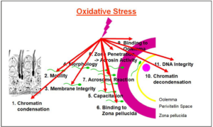
In sperm, oxidative stress is not only causing DNA fragmentation and mitochondrial dysfunction but is also negatively affecting gene expression and sperm-oocyte fusion [43-47]. Furthermore, oxidative stress can affect the genome and epigenome integrity [48,49]. In oocytes, the cytoskeleton, oocyte maturation and fertilization are negatively affected [50,51]. On the other hand, an excessive intake of antioxidants causes an opening of the disulfide bonds of protamines in the sperm head [52,53] and inhibit sperm capacitation [54]. Therefore, for normal reproductive functions of sperm and oocytes, the state of “oxidative eustress” with an approximate cellular H2O2 concentration between 1 nM and 10 nM H2O2 needs to be maintained [55], i.e. the maintenance of the cellular redox homeostasis (Figure 2).

Impact of redox stress on male and female fertility:

Male and female fertility can be compromised by numerous reproductive dysregulations, adverse lifestyle, aging, environmental pollutants or radio- and chemotherapy. In most of these conditions, oxidative stress is significantly involved as a major cause or contributor to the infertility. In men, a major cause of infertility is varicocele. While in the general population, this condition is found in 15% of the adult men, the prevalence is 35% and up to 80% in men with primary and secondary infertility, respectively, much higher [56-59]. Although all of these conditions have different aetiologies, oxidative stress has been implicated as a major mediator for reproductive dysfunctions, thus  infertility [60-68].

In sperm, oxidative stress specifically leads to nuclear and mitochondrial DNA decays, including DNA fragmentation [69,70], DNA methylation [71], and telomere changes and attrition [72,73], as well as and mitochondrial DNA damage resulting in mitochondrial dysfunction [74,75]. On the other hand, due to the extraordinary high susceptibility of sperm plasma membranes to oxidative assaults, this leads to lipid peroxidation of membrane lipids, resulting in poor motility, compromised acrosome reaction and the inability of sperm to fuse with the oolemma [76,77], thus poor fertilization ability. Similarly, on the female side, the oocytes, if exposed to excessive oxidative stress, sustain DNA damage including mtDNA damage, altered gene expression, mitochondrial dysfunction, impaired oocyte maturation and luteolysis [78-81].

Sperm health

Focusing on sperm health, functions and criteria, numerous studies have shown the detrimental effects of oxidative stress on all sperm functions [82-84], therefore drastically impairing male fertility (Figure 3). On the other hand, one must always consider that normal physiological functions require a small amount of ROS. Panner Selvam et al. [36], by determining the oxidation-reduction potential (ORP) using the MiOXSYS system, showed that good sperm motility, vitality, and normal mitochondrial membrane potential (MMP) depend on a physiological level of ROS [36]. If the redox potential is too high under oxidative stress conditions or too low under reductive stress conditions, sperm will not function properly anymore as indicated by decreased motility, vitality and MMP. The authors also showed that under oxidative and reductive stress conditions, the expression of 3 proteins of the mitochondrial OXPHOS complex, CV-ATPA, CIII-UQCRC2 and CIV-MTCO1 significantly decreased after exposure of sperm to oxidative and reductive stress conditions with CV-ATPA and CIII-UQCRC2 almost disappearing. Essentially, ROS produced in mitochondria of non-capacitating sperm may influence the development of the early embryo by inducing  sperm DNA fragmentation [85].

Oocyte and ovarian health

Redox homeostasis is also required for female fertility. Here, ROS are essential for the regulation of the ovarian cycle and ovarian steroidogenesis [87], including oocyte maturation [88], ovulation [89], follicle development, atresia and luteolysis [90,91]. It has also been shown that oxidative stress is intimately involved in ovarian aging, mitochondrial dysfunction, spindle abnormalities, telomere shortening and inflammatory processes such as PCOS or endometriosis [92-95]. In these syndromes, oxidative stress with subsequent mitochondrial dysfunction can affect folliculogenesis, the dialogue between oocyte and cumulus cells, maturation of the ooplasm, chromosome segregation by increasing aneuploidy risks, and cause atresia of granulosa and theca cells. In order to support healthy oocyte development and quality, follicular fluid contains high amounts of various antioxidants [96,97]. Among the antioxidant enzymes, superoxide dismutase [98], catalase [99] and glutathione [100,101] are critical for the maintenance of redox homeostasis for ovulation, the luteal phase, for the protection of genomic integrity, or the development and protection of oocytes.

In follicular fluid of older women, Carbone and co-workers found lower activities of glutathione and catalase together with a lower ratios of catalase/superoxide dismutase and glutathione peroxidase/superoxide dismutase [96]. Age-related oxidative damage as observed as lipid peroxidation, damage to proteins and DNA has also been reported in follicles and ovarian tissue of older women [102,103], thus causing cellular dysfunction, affecting follicle development and consequently oocyte quality [104,105]. This increase in oxidative stress is not only seen in aging women, but also in infertile ones. Zaha et al. [106] reported significantly higher levels of free glutathione in follicular fluid of infertile women as compared to a fertile control group while other oxidative stress markers such as malondialdehyde or superoxide dismutase showed no difference [106]. Also, none of the oxidative stress markers investigated was affected in serum. The higher glutathione levels in follicular fluid of infertile women could possibly be explained as a counter-regulation mechanism against oxidative stress. Furthermore, since oxidative stress is resulting in mitochondrial dysfunction with altered mitochondrial membrane potential, this leads to a release of cytochrome c triggering apoptosis [107]. Since telomeres are very prone to oxidation and due to heterochromatin state of telomeres, these damages are less efficiently repaired [108,109]. A study by Yamada-Fukunaga et al. [110] showed that telomeres in oocytes from older females are significantly shorter.

Effect of carnitines

Aside from eliminating the root cause of oxidative stress, treating it with antioxidants is one of the options. Among the different antioxidants that can neutralize high ROS levels causing oxidative stress is L-carnitine (β-hydroxy-γ-trimethylaminobutyrate), a natural antioxidant that is produced in living organisms from the amino acids lysine and methionine in the liver and kidney. While about 25% of the L-carnitine is synthesized endogenously, the remaining 75% have to be provided by food [7]. L-Carnitine is found in large quantities in red meat, especially mutton and lamb whereas poultry has less L-carnitine and vegetarian foods very little or no L-carnitine.

In human cells, L-carnitine has vital roles in alleviating cellular damage by transferring long-chain fatty acids to the internal mitochondrial membrane for β-oxidation, modulating acyl-Co-A/CoA, reducing the acyl group toxicity by excreting carnitine esters, and having antioxidant and antiradical properties [7,111,112]. L-carnitine improves mitochondrial functions by shuttling fatty acids into β-oxidation, thereby preventing accumulation of damaged lipids [113,114], hence preventing lipid peroxidation of the sensitive sperm membranes. With regard to sperm, oocytes and embryo development, L-carnitine has been shown to improve sperm count, motility, morphology and DNA fragmentation, and regulate oocyte and embryo energetics [115-118] and can therefore improve fertility rates. Treatment of infertile women with L-carnitine resulted in significantly higher transferable embryo rates as well as better quality of embryos [119]. In a systematic review and meta-analysis of 69 studies, Zafar and co-workers reported significantly higher pregnancy rates where men were supplemented with L-carnitine and micronutrients [120]. Similarly, in a randomized, double-blind, placebo-controlled trial including 263 patients, Lahimer et al. [121] found that supplementation of infertile men with L-carnitine and micronutrients significantly improved clinical pregnancy and live birth rates.

Conclusion

In conclusion, mitochondria are the site in eukaryotes where the required energy as well as ROS are produced and are crucial for sperm and oocyte development and function. Although small amounts of ROS are necessary to trigger essential cellular functions, excessive amounts of ROS that are not counteracted by a sufficient amount of antioxidants cause diseases and negatively affect sperm and oocyte functions, hence fertilization and embryo development. Therefore, maintaining redox homeostasis is essential for normal reproductive functions in men and women. In case of oxidative stress due to a number of diseases and health and lifestyle conditions, supplementation with antioxidants can improve the situation. Among these, L-carnitine has shown to be of particular importance as this quaternary amine has not only direct antioxidant properties but is also intimately involved in shuttling long-chain fatty acids into the mitochondrial β-oxidation for energy production as well as the detoxification of damaging lipids.

Acknowledgement

This document was a lecture presented by R.H, at the 15th Congress of the European Society of Gynecology, November 29 to December 2, 2023, Amsterdam, The Netherlands.

Conflicts of interest

The author declares having no conflicts of interest.

Funding

None.

Media

References

  1. Margulis L. Origin of eukaryotic cells. 1970. Yale University Press, New Haven, CT.
  2. Ankel-Simons F, Cummins JM. Misconceptions about mitochondria and mammalian fertilization: implications for theories on human evolution. Proc Natl Acad Sci U S A. 1996;93(24):13859-13863.
  3. Mortimer D. The functional anatomy of the human spermatozoon: relating ultrastructure and function. Mol Hum Reprod. 2018;24(12):567-592.
  4. Cummins JM. Mitochondria: potential roles in embryogenesis and nucleocytoplasmic transfer. Hum Reprod Update. 2001;7(2):217-228.
  5. Fisher D, Henkel R. Mitochondrial function and male infertility. In: Arafa M, ElBardisi H, Majzoub A, Agarwal A (Eds.). Genetics of Male Infertility. A Case-Based Guide for Clinicians. 2020; Springer Nature, Cham, Switzerland. pp: 137-153.
  6. Ferramosca A, Zara V. Bioenergetics of mammalian sperm capacitation. Biomed Res Int. 2014;2014:902953.
  7. Longo N, Frigeni M, Pasquali M. Carnitine transport and fatty acid oxidation. Biochim Biophys Acta. 2016;1863(10):2422-2435.
  8. Spinelli JB, Haigis MC. The multifaceted contributions of mitochondria to cellular metabolism. Nat Cell Biol. 2018;20(7):745-754.
  9. Boveris A, Chance B. The mitochondrial generation of hydrogen peroxide. General properties and effect of hyperbaric oxygen. Biochem J. 1973;134(3):707-716.
  10. Chance B, Sies H, Boveris A. Hydroperoxide metabolism in mammalian organs. Physiol Rev. 1979;59(3):527-605.
  11. Scialò F, Fernández-Ayala DJ, Sanz A. Role of mitochondrial reverse electron transport in ROS signaling: Potential roles in health and disease. Front Physiol. 2017;8:428.
  12. Halliwell B, Gutteridge JMC. Free Radicals in Biology and Medicine, 2nd Oxford University Press. 1989. Oxford, UK.
  13. Raha S, Robinson BH. Mitochondria, oxygen free radicals, disease and ageing. Trends Biochem Sci. 2000;25(10):502-508.
  14. Luo G, Ono S, Beukes NJ, Wang DT, Xie S, Summons RE. Rapid oxygenation of Earth’s atmosphere 2.33 billion years ago. Sci Adv. 2016;2(5):e1600134.
  15. Brochier-Armanet C, Talla E, Gribaldo S. The multiple evolutionary histories of dioxygen reductases: Implications for the origin and evolution of aerobic respiration. Mol Biol Evol. 2009;26(2):285-297.
  16. Marinho HS, Real C, Cyrne L, Soares H, Antunes F. Hydrogen peroxide sensing, signaling and regulation of transcription factors. Redox Biol. 2014;2:535-562.
  17. Allen RG, Tresini M. Oxidative stress and gene regulation. Free Radic Biol Med. 2000;28(3):463-499.
  18. Banerjee Mustafi S, Chakraborty PK, Dey RS, Raha S. Heat stress upregulates chaperone heat shock protein 70 and antioxidant manganese superoxide dismutase through reactive oxygen species (ROS), p38MAPK, and Akt. Cell Stress Chaperones. 2009;14(6):579-589.
  19. Massaad CA, Klann E. Reactive oxygen species in the regulation of synaptic plasticity and memory. Antioxid Redox Signal. 2011;14(10):2013-2054.
  20. Tschopp J. Mitochondria: Sovereign of inflammation? Eur J Immunol. 2011;41(5):1196-1202.
  21. Sies H, Belousov VV, Chandel NS, et al. Defining roles of specific reactive oxygen species (ROS) in cell biology and physiology. Nat Rev Mol Cell Biol. 2022;23(7):499-515.
  22. Gray MW. Mitochondrial evolution. Cold Spring Harb Perspect Biol. 2012;4(9):a011403.
  23. Folgerø T, Bertheussen K, Lindal S, Torbergsen T, Oian P. Mitochondrial disease and reduced sperm motility. Hum Reprod. 1993;8(11):1863-1868.
  24. Pesole G, Gissi C, De Chirico A, Saccone C. Nucleotide substitution rate of mammalian mitochondrial genomes. J Mol Evol. 1999;48(4):427-434.
  25. Murphy MP. Modulating mitochondrial intracellular location as a redox signal. Sci Signal. 2012;5(242):pe39.
  26. Caston RA, Demple B. Risky repair: DNA-protein crosslinks formed by mitochondrial base excision DNA repair enzymes acting on free radical lesions. Free Radic Biol Med. 2017;107:146-150.
  27. Yang SG, Park HJ, Kim JW, et al. Mito-TEMPO improves development competence by reducing superoxide in preimplantation porcine embryos. Sci Rep. 2018;8(1):10130.
  28. Lord T, Martin JH, Aitken RJ. Accumulation of electrophilic aldehydes during postovulatory aging of mouse oocytes causes reduced fertility, oxidative stress, and apoptosis. Biol Reprod. 2015;92(2):33.
  29. Aitken RJ, Whiting S, De Iuliis GN, McClymont S, Mitchell LA, Baker MA. Electrophilic aldehydes generated by sperm metabolism activate mitochondrial reactive oxygen species generation and apoptosis by targeting succinate dehydrogenase. J Biol Chem. 2012;287(39):33048-33060.
  30. Sies H. Oxidative Stress: Introductory Remarks. In: Sies E (Ed.), 1985. Oxidative Stress. Academic Press, London. pp:1-8.
  31. Sies H. Oxidative stress: oxidants and antioxidants. Exp Physiol. 1997;82(2):291-295.
  32. Zhang H, Limphong P, Pieper J, et al. Glutathione-dependent reductive stress triggers mitochondrial oxidation and cytotoxicity. FASEB J. 2012;26(4):1442-1451.
  33. Lloret A, Fuchsberger T, Giraldo E, Vina J. Reductive stress: A new concept in Alzheimer’s disease. Curr Alzheimer Res. 2016;13(2):206-211.
  34. Peris E, Micallef P, Paul A, et al. Antioxidant treatment induces reductive stress associated with mitochondrial dysfunction in adipocytes. J Biol Chem. 2019;294(7):2340-2352.
  35. Ma WX, Li CY, Tao R, Wang XP, Yan LJ. Reductive stress-induced mitochondrial dysfunction and cardiomyopathy. Oxid Med Cell Longev. 2020;2020:5136957.
  36. Panner Selvam MK, Agarwal A, Henkel R, et al. The effect of oxidative and reductive stress on semen parameters and functions of physiologically normal human spermatozoa. Free Radic Biol Med. 2020;152:375-385.
  37. Castagné V, Lefèvre K, Natero R, Clarke PG, Bedker DA. An optimal redox status for the survival of axotomized ganglion cells in the developing retina. Neuroscience. 1999;93(1):313-320.
  38. Lushchak VI, Storey KB. Oxidative stress concept updated: Definitions, classifications, and regulatory pathways implicated. EXCLI J. 2021;20:956-967.
  39. Finkel T, Holbrook NJ. Oxidants, oxidative stress and the biology of ageing. Nature. 2020;408(6809):239-247.
  40. Chrousos GP. Stress and disorders of the stress system. Nat Rev Endocrinol. 2009;5(7):374-381.
  41. Mentor S, Fisher D. Aggressive antioxidant reductive stress impairs brain endothelial cell angiogenesis and blood brain barrier function. Curr Neurovasc Res. 2017;14(1):71-81.
  42. Henkel R, Sandhu IS, Agarwal A. The excessive use of antioxidant therapy: A possible cause of male infertility? Andrologia. 2019;51(1):e13162.
  43. Aitken RJ, Gordon E, Harkiss D, et al. Relative impact of oxidative stress on the functional competence and genomic integrity of human spermatozoa. Biol Reprod. 1998;59(5):1037-1046.
  44. Gonçalves FS, Barretto LSS, Arruda RP, Perri SHV, Mingoti GZ. Effect of antioxidants during bovine in vitro fertilization procedures on spermatozoa and embryo development. Reprod Domest Anim. 2010;45(1):129-135.
  45. Takahashi M. Oxidative stress and redox regulation on in vitro development of mammalian embryos. J Reprod Dev. 2012;58(1):1-9.
  46. Muratori M, Tamburrino L, Marchiani S, et al. Investigation on the origin of sperm DNA fragmentation: Role of apoptosis, immaturity and oxidative stress. Mol Med. 2015;21(1):109-122.
  47. Aitken RJ. Impact of oxidative stress on male and female germ cells: implications for fertility. Reprod. 2020; 159(4):R189–R201.
  48. Dada R, Kumar SB, Chawla B, Bisht S, Khan S. Oxidative stress induced damage to paternal genome and impact of meditation and Yoga – Can it reduce incidence of childhood cancer? Asian Pac J Cancer Prev. 2016;17(9):4517-4525.
  49. Sudhakaran G, Kesavan D, Kandaswamy K, Guru A, Arockiaraj J. Unravelling the epigenetic impact: Oxidative stress and its role in male infertility-associated sperm dysfunction. Reprod Toxicol. 2024;124:108531.
  50. Combelles CM, Gupta S, Agarwal A. Could oxidative stress influence the in-vitro maturation of oocytes? Reprod Biomed Online. 2009;18(6):864-880.
  51. Lin J, Wang L. Oxidative stress in oocytes and embryo development: Implications for in vitro systems. Antioxid Redox Signal. 2020 Dec 8. doi: 10.1089/ars.2020.8209. Online ahead of print.
  52. Menezo YJ, Hazout A, Panteix G, et al. Antioxidants to reduce sperm DNA fragmentation: an unexpected adverse effect. Reprod Biomed Online. 2007;14(4):418-421.
  53. Giustarini D, Dalle-Donne I, Colombo R, Milzani A, Rossi R. Is ascorbate able to reduce disulfide bridges? A cautionary note. Nitric Oxide. 2008;19(3):252-258.
  54. de Lamirande E, Eiley D, Gagnon C. Inverse relationship between the induction of human sperm capacitation and spontaneous acrosome reaction by various biological fluids and the superoxide scavenging capacity of these fluids. Int J Androl. 1993;16(4):258-266.
  55. Sies H, Jones DP. Reactive oxygen species (ROS) as pleiotropic physiological signalling agents. Nat Rev Mol Cell Biol. 2020;21(7):363-383.
  56. Dubin L, Amelar RD. Etiologic factors in 1294 consecutive cases of male infertility. Fertil Steril. 1971;22(8):469-474.
  57. Masson P, Brannigan RE. The varicocele. Urol Clin North Am. 2014;41(1):129-144.
  58. Alsaikhan B, Alrabeeah K, Delouya G, Zini A. Epidemiology of varicocele. Asian J Androl. 2016;18(2):179-181.
  59. Jungwirth A, Diemer T, Kopa Z, Krausz C, Minhas S, Tournaye H. EAU Guidelines on Male Infertility. Edition. Presented at the EAU Annual Congress Barcelona 2019. ISBN 978-94-92671-04-2.
  60. Morris ID. Sperm DNA damage and cancer treatment. Int J Androl. 2002;25(5):255-261.
  61. Sakamoto Y, Ishikawa T, Kondo Y, Yamaguchi K, Fujisawa M. The assessment of oxidative stress in infertile patients with varicocele. BJU Int. 2008;101(12):1547-1552.
  62. Gupta S, Fedor, J, Biedenharn K, Agarwal A. Lifestyle factors and oxidative stress in female infertility: is there an evidence base to support the linkage? Expert Rev Obstet Gynecol. 2013;8(6):607–624.
  63. Murri M, Luque-Ramírez M, Insenser M, Ojeda-Ojeda M, Escobar-Morreale HF. Circulating markers of oxidative stress and polycystic ovary syndrome (PCOS): a systematic review and meta-analysis. Hum Reprod Update. 2013;19(3):268-288.
  64. Wright C, Milne S, Leeson H. Sperm DNA damage caused by oxidative stress: modifiable clinical, lifestyle and nutritional factors in male infertility. Reprod Biomed Online. 2014;28(6):684-703.
  65. Agarwal A, Rana M, Qiu E, AlBunni H, Bui AD, Henkel R. Role of oxidative stress, infection and inflammation in male infertility. Andrologia. 2018;50(11):e13126.
  66. Nago M, Arichi A, Omura N, Iwashita Y, Kawamura T, Yumura Y. Aging increases oxidative stress in semen. Investig Clin Urol. 2021;62(2):233-238.
  67. Wang L, Tang J, Wang L, et al. Oxidative stress in oocyte aging and female reproduction. J Cell Physiol. 2021;236(12):7966-7983.
  68. Ansariniya H, Yavari A, Javaheri A, Zare F. Oxidative stress-related effects on various aspects of endometriosis. Am J Reprod Immunol. 2022;88(3):e13593.
  69. Lopes S, Jurisicova A, Sun JG, Casper RF. Reactive oxygen species: potential cause for DNA fragmentation in human spermatozoa. Hum Reprod. 1998; 13(4):896-900.
  70. Henkel R, Hajimohammad M, Stalf T, et al. Influence of deoxyribonucleic acid damage on fertilization and pregnancy. Fertil Steril. 2004;81(4):965-972.
  71. Aston KI, Uren PJ, Jenkins TG, et al. Aberrant sperm DNA methylation predicts male fertility status and embryo quality. Fertil Steril. 2015;104(6):1388-1397.
  72. Bui AD, Sharma R, Henkel R, Agarwal A. Reactive oxygen species impact on sperm DNA and its role in male infertility. Andrologia. 2018;50(8):e13012.
  73. M’kacher R, Colicchio B, Marquet V, et al. Telomere aberrations, including telomere loss, doublets, and extreme shortening, are increased in patients with infertility. Fertil Steril. 2021;115(1):164-173.
  74. Cassina A, Silveira P, Cantu L, Montes JM, Radi R, Sapiro R. Defective human sperm cells are associated with mitochondrial dysfunction and oxidant production. Biol Reprod. 2015;93(5):119.
  75. Durairajanayagam D, Singh D, Agarwal A, Henkel R. Causes and consequences of sperm mitochondrial dysfunction. Andrologia. 2020;53(1):e13666.
  76. Baumber J, Ball BA, Gravance CG, Medina V, Davies-Morel MC. The effect of reactive oxygen species on equine sperm motility, viability, acrosomal integrity, mitochondrial membrane potential, and membrane lipid peroxidation. J Androl. 2000;21(6):895-902.
  77. Moawad AR, Fernandez MC, Scarlata E, et al. Deficiency of peroxiredoxin 6 or inhibition of its phospholipase A2 activity impair the in vitro sperm fertilizing competence in mice. Sci Rep. 2017;7(1):12994.
  78. Menezo YJ, Silvestris E, Dale B, Elder K. Oxidative stress and alterations in DNA methylation: two sides of the same coin in reproduction. Reprod Biomed Online. 2016; 33(6):668-683.
  79. Cacciottola L, Donnez J, Dolmans MM. Oxidative stress, mitochondria, and infertility: Is the relationship fully established? Fertil Steril. 2021;116(2):306-308.
  80. Hussain T, Murtaza G, Metwally E, et al. The role of oxidative stress and antioxidant balance in pregnancy. Mediators Inflamm. 2021;2021:9962860.
  81. Kordowitzki P. Oxidative stress induces telomere dysfunction and shortening in human oocytes of advanced age Donors. Cells. 2021;10(8):1866.
  82. Sikka SC. Oxidative stress and role of antioxidants in normal and abnormal sperm function. Front Biosci. 1996;1:e78-86.
  83. Chen SJ, Allam JP, Duan YG, Haidl G. Influence of reactive oxygen species on human sperm functions and fertilizing capacity including therapeutical approaches. Arch Gynecol Obstet. 2013;288(1):191-199.
  84. Dutta S, Majzoub A, Agarwal A. Oxidative stress and sperm function: A systematic review on evaluation and management. Arab J Urol. 2019;17(2):87-97.
  85. Mateo-Otero Y, Llavanera M, Torres-Garrido M, Yeste M. Embryo development is impaired by sperm mitochondrial-derived ROS. Biol Res. 2024;57(1):5.
  86. Henkel R. Leukocytes and oxidative stress: dilemma for sperm function and male fertility. Asian J Androl. 2011;13(1):43-52.
  87. Sawada M, Carlson JC. Intracellular regulation of progesterone secretion by the superoxide radical in the rat corpus luteum. Endocrinology. 1996;137(5):1580-1584.
  88. Miyazaki T, Sueoka K, Dharmarajan AM, Atlas SJ, Bulkley GB, Wallach EE. Effect of inhibition of oxygen free radical on ovulation and progesterone production by the in-vitro perfused rabbit ovary. J Reprod Fertil. 1991;91(1):207-212.
  89. Shkolnik K, Tadmor A, Ben-Dor S, Nevo N, Galiani D, Dekel N. Reactive oxygen species are indispensable in ovulation. Proc Natl Acad Sci USA. 2011;108(4):1462-1467.
  90. Sugino N. Reactive oxygen species in ovarian physiology. Reprod Med Biol. 2005;4(1):31-44.
  91. Sugino N. Roles of reactive oxygen species in the corpus luteum. Anim Sci J. 2006;77:556-565.
  92. Sasaki H, Hamatani T, Kamijo S, et al. Impact of oxidative stress on age-associated decline in oocyte developmental competence. Front Endocrinol (Lausanne). 2019;10:811.
  93. Zhang J, Bao Y, Zhou X, Zheng L. Polycystic ovary syndrome and mitochondrial dysfunction. Reprod Biol Endocrinol. 2019;17(1):67.
  94. Yang L, Chen Y, Liu Y, et al. The role of oxidative stress and natural antioxidants in ovarian aging. Front Pharmacol. 2021;11:617843.
  95. Fan W, Yuan Z, Li M, Zhang Y, Nan F. Decreased oocyte quality in patients with endometriosis is closely related to abnormal granulosa cells. Front Endocrinol (Lausanne). 2023;14:1226687.
  96. Carbone MC, Tatone C, Delle Monache S, et al. Antioxidant enzymatic defences in human follicular fluid: characterization and age-dependent changes. Mol Hum Reprod. 2003;9(11):639-643.
  97. Wang S, He G, Chen M, Zuo T, Xu W, Liu X. The role of antioxidant enzymes in the ovaries. Oxid Med Cell Longev. 2017;2017:4371714.
  98. Sugino N, Takiguchi S, Kashida S, Karube A, Nakamura Y, Kato H. Superoxide dismutase expression in the human corpus luteum during the menstrual cycle and in early pregnancy. Mol Hum Reprod. 2000;6(1):19-25.
  99. Park YS, You SY, Cho S, et al. Eccentric localization of catalase to protect chromosomes from oxidative damages during meiotic maturation in mouse oocytes. Histochem Cell Biol. 2016;146(3):281-288.
  100. Maedomari N, Kikuchi K, Ozawa M, et al. Cytoplasmic glutathione regulated by cumulus cells during porcine oocyte maturation affects fertilization and embryonic development in vitro. Theriogenology. 2007;67(5):983-993.
  101. Hoang YD, Nakamura BN, Luderer U. Follicle-stimulating hormone and estradiol interact to stimulate glutathione synthesis in rat ovarian follicles and granulosa cells. Biol Reprod. 2009;81(4):636-646.
  102. Lim J, Luderer U. Oxidative damage increases and antioxidant gene expression decreases with aging in the mouse ovary. Biol Reprod. 2011;84(4):775-782.
  103. Zhang D, Zhang X, Zeng M, et al. Increased DNA damage and repair deficiency in granulosa cells are associated with ovarian aging in rhesus monkey. J Assist Reprod Genet. 2015;32(7):1069-1078.
  104. González-Fernández R, Hernández J, Martín-Vasallo P, Puopolo M, Palumbo A, Ávila J. Expression levels of the oxidative stress response gene ALDH3A2 in granulosa-lutein cells are related to female age and infertility diagnosis. Reprod Sci. 2016;23(5):604-609.
  105. Timóteo-Ferreira F, Abreu D, Mendes S, et al. Redox imbalance in age-related ovarian dysfunction and perspectives for its prevention. Ageing Res Rev. 2021;68:101345.
  106. Zaha I, Muresan M, Tulcan C, et al. The role of oxidative stress in infertility. J Pers Med. 2023;13(8):1264.
  107. Redza-Dutordoir M, Averill-Bates DA. Activation of apoptosis signalling pathways by reactive oxygen species. Biochim Biophys Acta. 2016;1863(12):2977-2992.
  108. Smith S. Telomerase can’t handle the stress. Genes Dev. 2018;32(9-10):597-599.
  109. Singh A, Kukreti R, Saso L, Kukreti S. Oxidative stress: Role and response of short guanine tracts at genomic locations. Int J Mol Sci. 2019;20(17):4258.
  110. Yamada-Fukunaga T, Yamada M, Hamatani T, et al. Age-associated telomere shortening in mouse oocytes. Reprod Biol Endocrinol. 2013;11:108.
  111. Bremer J. Carnitine–metabolism and functions. Physiol Rev. 1983;63(4):1420-1480.
  112. Vanella A, Russo A, Acquaviva R, et al. L-propionyl-carnitine as superoxide scavenger, antioxidant, and DNA cleavage protector. Cell Biol Toxicol. 2000;16(2):99-104.
  113. Thangasamy T, Jeyakumar P, Sittadjody S, Joyee AG, Chinnakannu P. L-carnitine mediates protection against DNA damage in lymphocytes of aged rats. Biogerontology. 2009;10(2):163-172.
  114. Cabral REL, Mendes TB, Vendramini V, Miraglia SM. Carnitine partially improves oxidative stress, acrosome integrity, and reproductive competence in doxorubicin-treated rats. Andrology. 2018;6(1):236-246.
  115. Nezhad NC, Vahabzadeh Z, Allahveisie A, et al. The effect of L-carnitine and coenzyme Q10 on the sperm motility, DNA fragmentation, chromatin structure and oxygen free radicals during, before and after freezing in oligospermia men. Urol J. 2021;18(3):330-336.
  116. Li J, Liu L, Weng J, Yin TL, Yang J, Feng HL. Biological roles of l-carnitine in oocyte and early embryo development. Mol Reprod Dev. 2021;88(10):673-685.
  117. Placidi M, Di Emidio G, Virmani A, et al. Carnitines as mitochondrial modulators of oocyte and embryo bioenergetics. Antioxidants (Basel). 2022;11(4):745.
  118. Sun Y, Ma L. Comparison of L-carnitine vs. Coq10 and vitamin E for idiopathic male infertility: a randomized controlled trial. Eur Rev Med Pharmacol Sci. 2022;26(13):4698-4704.
  119. Kitano Y, Hashimoto S, Matsumoto H, et al. Oral administration of l-carnitine improves the clinical outcome of fertility in patients with IVF treatment. Gynecol Endocrinol. 2018;34(8):684-688.
  120. Zafar MI, Mills KE, Baird CD, Jiang H, Li H. Effectiveness of nutritional therapies in male factor infertility treatment: A systematic review and network meta-analysis. Drugs. 2023;83(6):531-546.
  121. Lahimer M, Gherissi O, Ben Salem N, et al. Effect of micronutrients and L-carnitine as antioxidant on sperm parameters, genome integrity, and ICSI outcomes: Randomized, double-blind, and placebo-controlled clinical trial. Antioxidants (Basel). 2023;12(11):1937.

Keywords: , , , ,

Citation: Henkel R., Role of mitochondria and redox state in sperm and oocyte health, GREM Gynecological and Reproductive Endocrinology & Metabolism (2024); 01/2024:027-033 doi: 10.53260/grem.2450105

Published: April 17, 2024